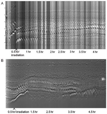
Figure 3. Kymograph of a centrosome irradiated cell and cytoplasm irradiated cell.
(A) Initially we observe forward movement of both the leading edge (top) and trailing edge (bottom) as determined by the incline formed by the cell edges. After the first 30 minute period, the cell was repositioned and the centrosome was irradiated by the laser. Following laser exposure we see no forward movement of the leading edge as determined by the horizontal line formed by the leading edge (top). At the trailing edge (bottom) we see a spreading of the cell edge forming a declining line. By the end of the 4.5 hour observation period, the cell front and back appear similar to each other and equally spaced from the nucleus. See Movie S1. (B) Over time, we see the cell constantly changing its shape and position. Regardless of its shape change, there is always a visible lamellae with a majority of the cytoplasm in front of the nucleus (in the direction of migration). See Movie S2.

