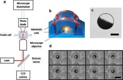
Figure 1.
(a) Schematic representation of the laser and microscope setup in which a low power laser, in conjunction with a dichroic mirror, a microscope objective and a photodiode, was used to measure the rotation rate of a single magnetic bead. A digital camera can be used to simultaneously capture a video of the rotating system. (b) Custom designed Helmholtz coils were used to create a rotating magnetic field in the imaging plane. (c) An optical microscope image of a half coated 10 μm bead (300 nm Nickel coating) with a 5 μm scale bar. (d) Image sequence of a 6.7 μm bead rotating synchronously in the LiveCell Array in a 10 Hz field. The time between each frame is 14 ms and the scale bar is 10 μm.

