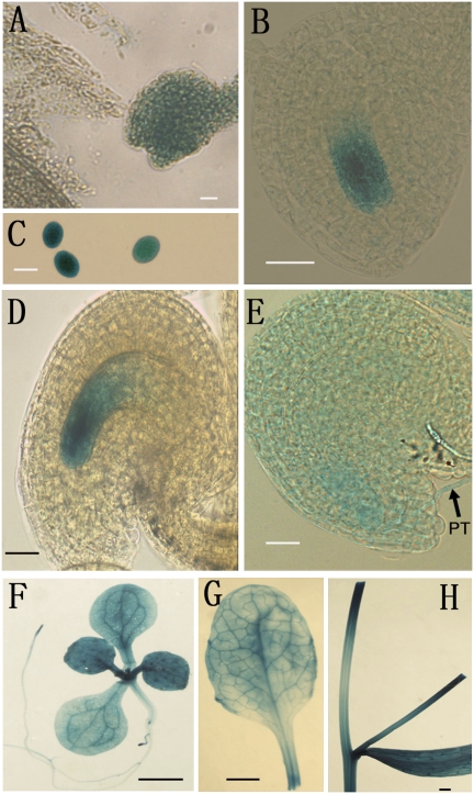
Fig. 1.
Expression patterns of ANK6 by GUS reporter analysis. Histochemical GUS staining of ovules from plants transformed with ANK6 promoter-GUS. GUS activities (indicated in blue) were observed at different developmental stages of male (C) and female (A, B, D, and E) gametophytes. GUS signals were also detectable in leaves (F and G), roots (F), and stems (H). Early one-nucleate embryo sac (A), FG1 stage (B), mature pollen (C), mature ovule (D), ovule with penetrating pollen tube (PT, arrow) (E), 2-wk-old plant (F), mature leaf (G), and mature stem (H) are shown. (Scale bars: A–E, 20 μm; F–H, 0.5 cm.)

