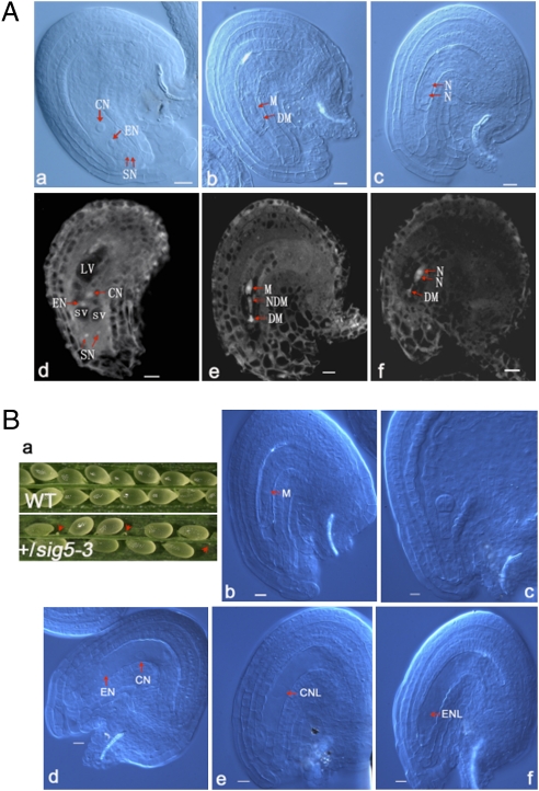
Fig. 6.
Ovules are arrested in early developmental stages in both ank6 and sig5 mutants. (A) Ovule development in WT plants and ank6 mutant revealed by Nomarski microscopy and CLSM. CN, central nucleus; DM, degenerated megaspore; EN, egg nucleus; LV, large vacuole; N, nucleate; NDM, nondegenerated megaspore other than the functional megaspore; SN, synergid nuclei; SV, small vacuole. Mature WT ovule with a four-celled embryo sac at FG7 (A, a and d), ank6 mutant ovule arrested at FG1 (A, b and e), and ank6 mutant ovule arrested at FG2 (A, c and f). (Scale bars: 10 μm.) (B) Ovule development in WT plants and in sig5 mutant revealed by Nomarski microscopy. CN, central nucleus; CNL, central nuclear-like nucleus; EN, egg nucleus; ENL, egg nuclear-like nucleus; M, megaspore. Silique of self-pollinated +/sig5 plants showing frequently observed degenerating ovules indicated by arrowheads (B, a), sig5 mutant ovule arrested at FG1 (B, b), embryo at early globular stage of development in normal ovule of the same silique of control (B, c), sig5 mutant ovule arrested with egg cell and central cell unfertilized (B, d), sig5 mutant ovule arrested with a central cell-like nucleus remaining (B, e), and sig5 mutant ovule arrested with an egg cell-like nucleus remaining (B, f). (Scale bars: 10 μm.)

