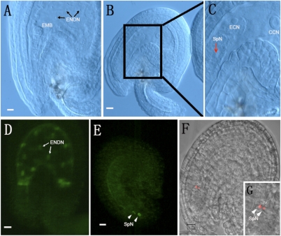
Fig. 7.
Assessment of gamete interaction in +/ank6 plants. Nomarski images of the ovules from a self-pollinated +/ank6 silique. CCN, central cell nucleus; ECN, egg cell nucleus; EMB, embryo; ENDN, endosperm nucleus; SpN, sperm nucleus. Morphologically normal ovule showing embryo at early globular stage of development (A), cleared mutant (unfertilized) ovule showing central cell, polarized egg cell (B and C), with sperm-like nucleus near egg cell in the sac (arrow). Paternally provided fluorescent LIG1-GFP marker is expressed after fertilization in the embryo and the endosperm. WT ovules and normal ovules in heterozygous plants developed from zygote elongation to 16-cell embryo stages with multiple endosperm nuclei (D; arrows); a small portion of the ovules in heterozygous plants are unfertilized with two sperm-like nuclei near the degenerated synergid cells (E; arrowheads). (F) Sperm-specific marker pHTR10-mRFP labeled free sperm nuclei after +/ank6 self-pollinated plants were examined. (G) Box shows the pHTR10-mRFP1 signals from two unfertilized sperm nuclei (arrowheads).

