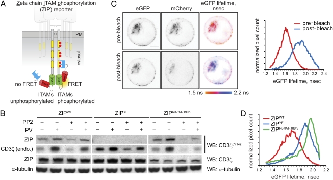
Fig. 1.
Design and characterization of the ZIP reporter. (A) Intramolecular binding of the SH2 domains to tyrosine-phosphorylated ITAMs within the ZIP reporter results in FRET between GFP and mCherry. (B) Jurkat cells stably expressing the ZIPWT reporter or the ZIPYF or ZIPR37K/R190K mutants were preincubated with the generic Src family kinase inhibitor PP2 (2 μM) or the vehicle for 30 min and then stimulated for 5 min with 100 μM sodium pervanadate (PV). Cell lysates were analyzed by Western blotting (WB) using mAb against CD3ζpY142, pan-CD3ζ, and α-tubulin. Full-length blots are presented in Fig. S1A. (C) Jurkat cells stably expressing the ZIPWT reporter were adhered onto glass coverslips precoated with 20 μg/mL anti-CD3 mAb HIT3a, and eGFP lifetime before and after photobleaching of the acceptor (mCherry) was monitored using TCSPC-FLIM. Shown are the confocal images in the eGFP and mCherry channels and the corresponding pseudocolored eGFP fluorescence lifetime images scaled between 1.5 and 2.2 ns. (Scale bar, 2 μm.) Right: Corresponding cumulative histograms of the eGFP lifetime before and after acceptor photobleaching. (D) Cumulative histograms of eGFP lifetime in Jurkat cells stably expressing the ZIPWT reporter or ZIPYF or ZIPR37K/R190K mutants, adhered onto glass coverslips precoated with 20 μg/mL HIT3 mAb for 10 min.

