Fig. 3.
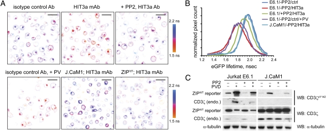
CD3ζ phosphorylation is controlled by a dynamic reaction cycle. (A) Wild-type Jurkat E6.1 or Lck-deficient J.CaM1 (Lower Center) cells stably expressing the ZIPWT reporter or the ZIPYF mutant (Lower Right) were preincubated with 2 μM PP2 or the vehicle (DMSO) for 30 min and then adhered onto a glass coverslip precoated with 20 μg/mL mAb HIT3a or the respective isotype control antibody for 10 min, fixed, and eGFP lifeime monitored using TCSPC-FLIM. Shown are pseudocolored eGFP lifetime images of typical regions of interest (ROIs), scaled between 1.5 and 2.2 ns. (Scale bars, 20 μm.) (B) Cumulative histograms of the eGFP lifetime averaged over 3 to 4 ROIs of cells stimulated as described for Fig. 2A. (C) Wild-type Jurkat E6.1 or Lck-deficient J.CaM1 cells stably expressing the ZIPWT reporter were preincubated with 2 μM PP2 or the vehicle (DMSO) for 30 min and then stimulated for 5 min with 100 μM sodium pervanadate. The lysates were analyzed by Western blotting using mAbs against CD3ζpY142, pan-CD3ζ, and α-tubulin. Full-length blots are presented in Fig. S2A.

