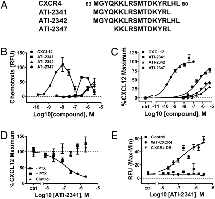
Fig. 1.
Identification and characterization of the CXCR4 agonist, ATI-2341. (A) Sequences of ATI-2341, 2342, and 2347. Depiction of the sequences of ATI-2341, ATI-2342, and ATI-2347 within the i1 loop region of human CXCR4. (B) ATI-2341, -2342, and -2347 dose-dependently elicit chemotaxis. Chemotaxis of CCRF-CEM cells was assessed in 96-well Transwell plates in response to a dose-range of CXCL12 or test articles in the bottom well. Cell migration was quantified using Cyquant dye. The averages of relative fluorescent intensity of duplicate wells were plotted. Data are representative of n = 3 independent experiments. (C) ATI-2341, -2342, and -2347 dose-dependently induce an increase in intracellular calcium in CCRF-CEM cells. CCRF-CEM cells preloaded with calcium 4 dye and incubated with varying concentrations of CXCL12 or test article and fluoresence intensity was recorded on a FlexStation III. Data were normalized to CXCL12-stimulated maximal calcium response. The averages of duplicate wells were plotted. Data are representative of n = 3 independent experiments. (D) ATI-2341 inhibits NKH477-stimulated cAMP in a CXCR4- and Gi-dependent manner. HEK-293 cells stably transfected with CXCR4, were pretreated (+PTX) or not pretreated (−PTX) with pertussis toxin overnight before the experiment. Control cells represent naïve HEK-293 cells. All cells were preincubated for 15 min with 10 μM of NKH477 before a 30-min incubation with ATI-2341. cAMP levels were then determined by HTRF assay. Data were normalized to the NKH477-stimulated intracellular cAMP level in the absence of compounds. The averages of triplicate wells were plotted. Data are representative of n = 3 independent experiments. (E) ATI-2341 stimulates calcium response in U87 cells transfected with human CXCR4 but not a loss-of-function mutant. U87 cells transfected with wild-type CXCR4, a CXCR4 with a mutation in the DRY motif to the sequence RDY (CXCR4-DR), or mock transfected were loaded with calcium 4 dye and incubated with varying levels of AT-2341. Fluoresence intensity was recorded on a FlexStation III. Data were normalized to CXCL12-stimulated maximal calcium response and the averages of triplicate wells were plotted. Data are representative of n = 3 independent experiments.

