Figure 6.
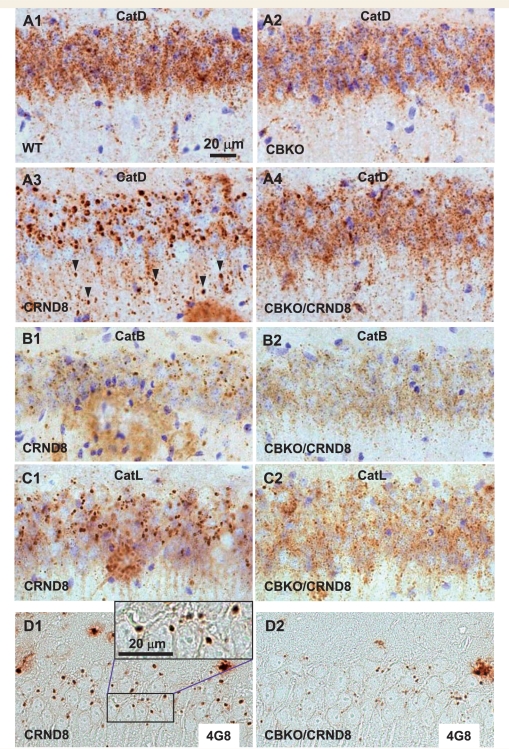
Elimination of giant autolysosomes in the brains of CBKO/TgCRND8. Vibratome (A–C) or paraffin (D) brain sections from a group of wild-type, CBKO, TgCRND8 and CBKO/TgCRND8 mice (strains: 129S6 × 129X1 for the four genotypes) at 6 months of age were processed with antibodies to cathepsin D (RU2) (A), cathepsin B (B), cathepsin L (C) or Aβ (4G8) (D). All images are from the hippocampal CA1 sector showing reduction of giant autolysosomes—revealed by antibodies to either cathepsins or Aβ—in the CBKO/TgCRND8 compared to TgCRND8. The images in (A) are representative of the results from 9–13 mice per genotype, while those in (B–D) are from 3–4 mice per genotype. Arrowheads (A3) depict giant autolysosomes within the stratum radiatum. Scale bars: 20 µm (A–D). CatB = cathepsin B; CatD = cathespin D; CatL = cathepsin L; CRND8 = TgCRND8.

