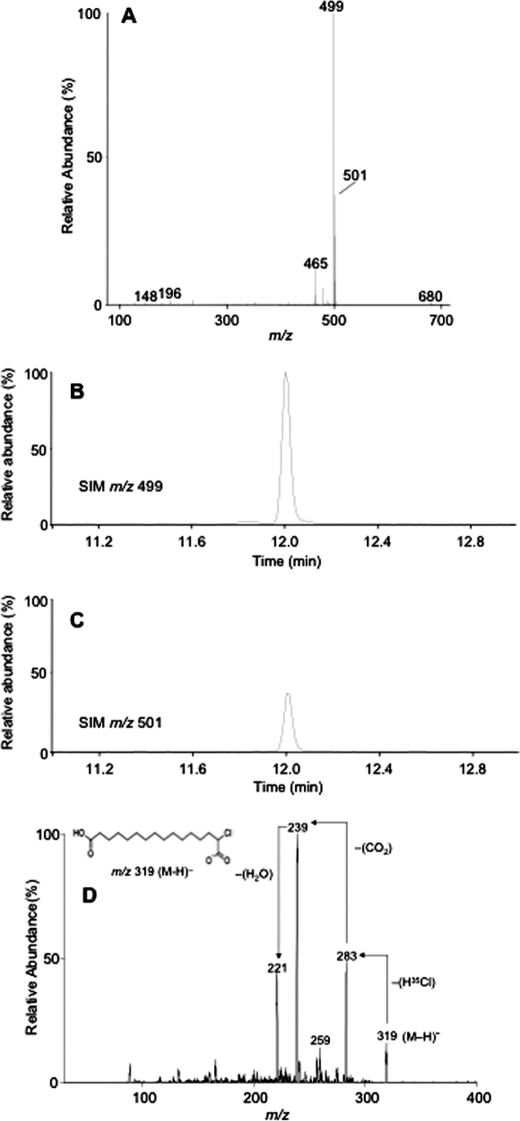
FIGURE 2.
Identification of 2-ClHDDA. HepG2 cells were incubated with 50 μm 2-ClHA for 24 h. At the end of the incubation period, media were extracted and analyzed by either GC-MS in the NICI mode following PFB derivatization (A–C) or by ESI-MS/MS of the underivatized extract (D). The mass spectrum of a peak obtained at 12 min, corresponding to the di-PFB ester of 2-ClHDDA, is depicted (A). The chromatograms obtained using SIM of m/z 499 (B) and m/z 501 (C) within a window of 11–12.2 min are shown. B and C, 100% relative abundances were set to equal ion counts. D shows the product-ion spectra of [M − H]− ion of 2-35ClHDDA. The prominent neutral losses (marked by arrows) of the proposed structure of the [M − H]− ion (inset) that is consistent with the starting material and the mass spectra are also shown.

