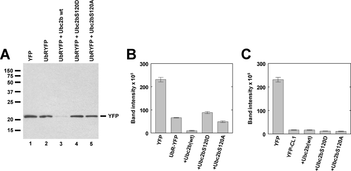
FIGURE 6.
Ser120 mutation inhibits degradation of a model N-end rule substrate. A, shown is a Western blot stained for YFP and visualized by chemiluminescence for pooled samples from triplicate plates of T47D human breast cancer cells transfected as described under “Materials and Methods” to ectopically express YFP or the model N-end rule substrate UbRYFP in the absence or presence of wild type or mutant HsUbc2b as indicated. Relative molecular weights are indicated to the left, and the migration position for free YFP is indicated to the right. B, shown is fluorescence quantitation of the Western blot of panel A. C, shown is fluorescence quantitation of the Western blot derived from a parallel experiment identical to that of panels A and B but expressing the model unfolded protein response substrate YFP-CL1. The YFP alone control is identical for panels A and B. Error bars correspond to the S.D. for triplicate samples.

