FIGURE 1.
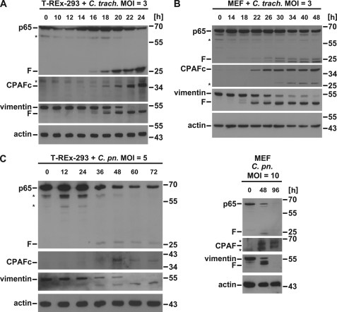
Infection with C. trachomatis causes cleavage of p65. A, C. trachomatis infection leads to p65 cleavage in T-REx-293 cells. Cells were infected with C. trachomatis with an m.o.i. of 3 for the times indicated. Cell lysates were subjected to Western blot analysis for p65, CPAF, or the CPAF substrate vimentin. Shown is a representative result of five independent experiments. F, cleavage products of p65; *, unspecific background bands. CPAFc, C-terminal autoprocessing product of CPAF. B, in MEF cells C. trachomatis infection leads to p65 cleavage. Mouse embryonic fibroblasts were infected with C. trachomatis with an m.o.i. of 3 as indicated. Radioimmune precipitation assay buffer extracts were prepared and subjected to Western blot analysis for p65, CPAF, or vimentin. Shown is a representative result of four independent experiments. C, C. pneumoniae infection causes p65 cleavage in both in T-REx-293 and MEF cells. Cells were infected with C. pneumoniae (m.o.i. of 5 for T-REx-293 or 10 for MEF) for the time periods indicated. Cell lysates were subjected to Western blot analysis for p65, CPAF, or the CPAF substrate vimentin. Shown is a representative result of three independent experiments.

