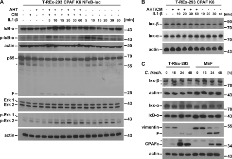
FIGURE 4.
NF-κB signaling upstream of p65/RelA remains functional during expression of active CPAF. A, phosphorylation of IκB-α upon stimulation with IL-1β is shown. T-REx-293 CPAF K6 NFκB-luc cells were treated with IL-1β (10 ng/ml) for the times indicated. In some aliquots of cells, active CPAF had been expressed by stimulation with AHT/CM for 7 h before the addition of IL-1β. Cells were lysed and subjected to Western blotting for IκB-α, phospho-IκB-α, Erk1/2 and phospho-Erk1/2. F, CPAF-specific cleavage products; *, unspecific background bands. Shown is a representative result of six independent experiments. B, IKK protein levels remain unchanged during expression of active CPAF. CPAF expressing cells (7 h after addition of AHT/CM) were treated with IL-1β (10 ng/ml) for the times indicated. Cell lysates were subjected to Western blotting. Shown is a representative result of five independent experiments. C, IKK protein levels remain unchanged after chlamydial infection. T-REx-293 cells or MEF cells were infected with C. trachomatis (m.o.i. = 3) for the times indicated. Cell extracts were analyzed by Western blotting. Shown is a representative result of three independent experiments.

