FIGURE 7.
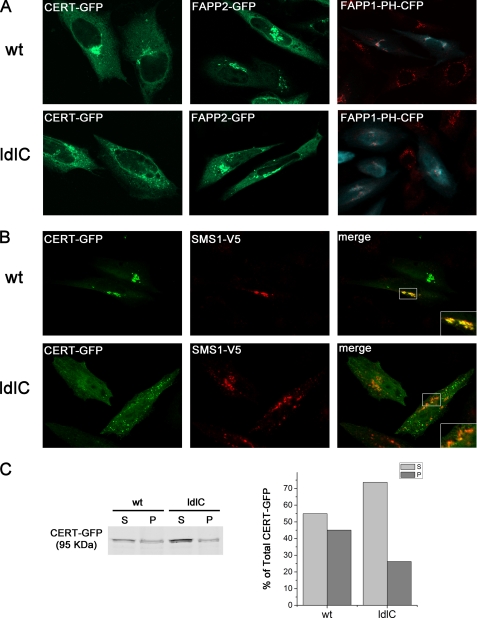
CERT, SMS1, FAPP2, and FAPP1-PH domain localization in WT and ldlC cells. A, CERT-GFP and FAPP1-PH-CFP but not FAPP2-GFP localization are altered in ldlC cells. WT and ldlC cells were transfected with CERT-GFP (green), FAPP2-GFP (green) or transfected wit FAPP1-PH-CFP (cyan) and stained for the endogenous Golgi market GM130 (red). Cells were fixed and observed under a confocal microscope. B, CERT and SMS1 localized to different membranes in ldlC cells. WT and ldlC cells were co-transfected with CERT-GFP and SMS1-V5. Cells were fixed with 3% paraformaldehyde in PBS for 20 min and permeabilized with Triton X-100 0.1% glycine (200 mm) for 10 min at room temperature and immunostained with monoclonal mouse anti-V5 and the appropriate secondary antibody conjugated with Alexa Fluor 546. WT cells show significant co-localization (Pearson's coefficient = 0.86 ± 0.05), whereas ldlC cells only show partial co-localization (Pearson's coefficient = 0.58 ± 0.04) between CERT and SMS1 in three independent experiments, with each analyzing 30 cells. Insets are a higher magnification of the boxed areas in the cells. C, WT and ldlC CERT-GFP-transfected cells were homogenized, and supernatant (S) and pellet (P) fractions were obtained by ultracentrifugation at 400,000 × g as described under “Experimental Procedures.”

