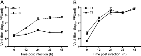
FIGURE 1.
Growth of reovirus in cultured cells. MDCK cells (A) or L cells (B) were infected at an m.o.i. of 2 pfu/cell, and viral titers in cell lysates were determined at the time points shown by plaque assay using L cell monolayers. Results represent the mean of triplicate experiments. Error bars indicate S.D.

