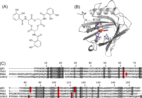
FIGURE 1.
Enterobactin and siderocalin(s). A, chemical structure of enterobactin. B, symbolic representation of the crystal structure of human NGAL in complex with FeDHBx (PDB accession code 1L6M), the Fe3+ ion is represented as a red sphere, the ligand moieties are depicted as blue sticks. C, sequence alignment of the secreted forms of putative siderocalins with NGAL; Q83: quail lipocalin Q83; Ch21: chicken lipocalin Ch21; NGAL: human NGAL; LCN12: human lipocalin 12, isoform a. Conserved and homologous residues are highlighted in dark and light gray, respectively, residues (putatively) involved in siderophore binding are highlighted in red.

