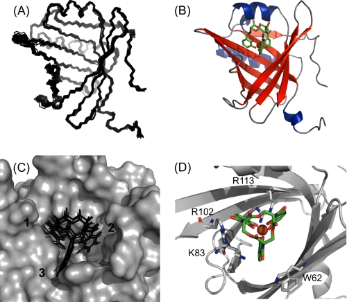
FIGURE 3.
Solution structure of the Q83/[GaIII(Ent)]3− complex. A, backbone superimposition of the final set of 20 structures for the Q83/[GaIII(Ent)]3− complex. B, representative ribbon model of the Q83/[GaIII(Ent)]3− complex. Enterobactin is represented as green sticks, Ga(III) is depicted as an orange sphere. C, ligand distribution within the Q83 calyx. The calyx surface of the most representative complex is represented in gray, enterobactin molecules are depicted as black sticks. D, structural details of the enterobactin binding site. Q83 residues involved in enterobactin binding are represented as gray sticks.

