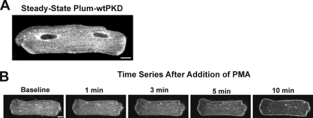
FIGURE 1.
Localization and movement of Plum-tagged wild type PKD (Plum-wtPKD) in myocytes activated with phorbol ester. ARVMs were infected with an adenovirus construct to drive expression of Plum-wtPKD and cultured for 24 h as described under “Experimental Procedures.” A, fluorescent confocal image of a single isolated ARVM that shows localization of Plum-wtPKD in the steady state. Scale bar, 10 μm. B, cells were stimulated with the phorbol ester derivative, PMA (2 nm), and a series of confocal images were acquired as indicated from the same cell after the addition of PMA.

