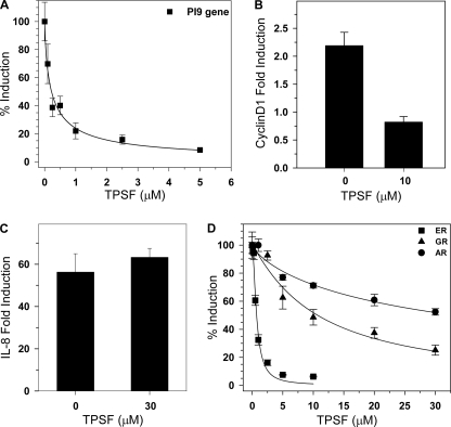
FIGURE 2.
TPSF specifically inhibits expression of endogenous ER-regulated genes. A, TPSF inhibits E2 induction of PI-9 mRNA. For studies of PI-9 mRNA (filled squares), MCF-7 cells were incubated for 24 h with the indicated concentrations of TPSF and maintained for 4 h in 10 nm E2 and TPSF. PI-9 mRNA was quantitated by RT-PCR as described (23). B, TPSF inhibition of E2-ERα induction of cyclin D1 mRNA is shown. MCF-7 cells were plated and 24 h later treated with ethanol and DMSO vehicles, 10 nm E2, or 10 nm E2 and 10 μm TPSF. After 24 h, RNA was extracted, and cyclin D1 mRNA levels were measured by qRT-PCR. The level of cyclin D1 mRNA in the vehicle only sample was set to 1. -Fold induction of cyclin D1 in the presence of 10 μm TPSF was significantly different from the control (p < 0.05 using Student's t test). C, TPSF does not inhibit NF-κB induction of IL-8 mRNA. MCF-7 cells were maintained for 24 h in medium without TNF-α or with 10 ng/ml TNF-α with and without 30 μm TPSF and harvested, and IL-8 mRNA levels were determined by qRT-PCR. D, dose-response studies of inhibition of ERα, AR, and GR transactivation are shown. For each receptor, induction of luciferase reporter gene expression (AR and GR) or endogenous PI-9 mRNA (ER) in the presence of an appropriate ligand with DMSO minus TPSF was set to 100%. Cells were incubated for 24 h with 0.2 nm E2 for ERα (filled squares), 5 nm dexamethasone for GR (filled triangles), 1 μm dihydrotestosterone for AR (filled circles), and the indicated concentrations of TPSF. Data are the average ± S.E. for three experiments.

