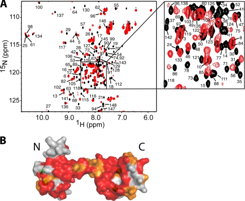
FIGURE 6.
A, overlay of two-dimensional 1H-15N HSQC spectra obtained for 15N-labeled CaM in the unbound state (black) and in complex with unlabeled myr(+)MA at 1. 4:1 (MA:CaM) (red). B, surface representation of the CaM structure of (Protein Data Base code 3CLN). Residues that exhibited substantial chemical shift changes (> 0.1 ppm) or signal loss are colored in red, whereas those modestly perturbed (< 0.1 ppm) are colored in orange. Calcium ions are colored in green.

