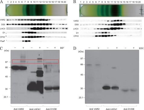
FIGURE 5.
Sucrose density gradient centrifugation and chemical cross-linking analysis showing the localization of FtsH proteases in the thylakoids and PSII membranes. A, sucrose density gradient centrifugation following the solubilization of thylakoids with DM. After centrifugation, the fractions of the tube were subjected to SDS/urea-PAGE and Western blot analysis with specific antibodies (indicated on the left-hand side of the gels). B, the same as A except PSII membranes were used as the sample. C, chemical cross-linking study of PSII membranes using BS3 to identify the nearest neighbor protein interactions. After the separation of the proteins in the PSII membranes by SDS/urea-PAGE, the antibodies against VAR2, LHCb1, and the DE loop of the D1 protein were used for the subsequent Western blot analysis. − and + at the top of the gel indicate the absence and presence of 0.25 mm BS3, respectively. The bands that appeared after the cross-linking reaction are indicated by the red square and represent the cross-linked products of FtsH and LHCb1. Molecular markers are shown on the left-hand side of the gel. D, chemical cross-linking study of PSII membranes using EDC. The concentration of EDC was 5 mm. The other conditions were the same as those described in C.

