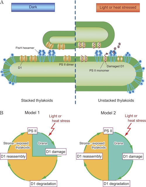
FIGURE 7.
A schematic representation of the distribution of FtsH proteases in thylakoid membranes and models showing turnover of the D1 protein. A, distribution of FtsH in thylakoid membranes. Hexameric FtsH proteases are abundant at the grana regions. Under light or heat stress, the damaged PSII dimers are converted to monomers, and degradation of the damaged D1 protein is initiated by the action of FtsH proteases that are present near the PSII complexes in the grana. In contrast, FtsH proteases with various subunit structures exist in the stroma thylakoids, and they probably represent the monomers, intermediate oligomers, and hexamers in the course of the assembly and degradation process of FtsH. Both the assembly and degradation of FtsH are possibly triggered by light and heat stresses. Under these conditions, the thylakoids show swelling and unstacking, which may stimulate the interaction between FtsH proteases and damaged PSII complexes. B, two models depicting turnover of the D1 protein under light or heat stress. Model 1 indicates that degradation of the D1 protein occurs exclusively in the unstacked regions of thylakoids, whereas Model 2 predicts that D1 degradation takes place both in the grana and the unstacked regions of thylakoids.

