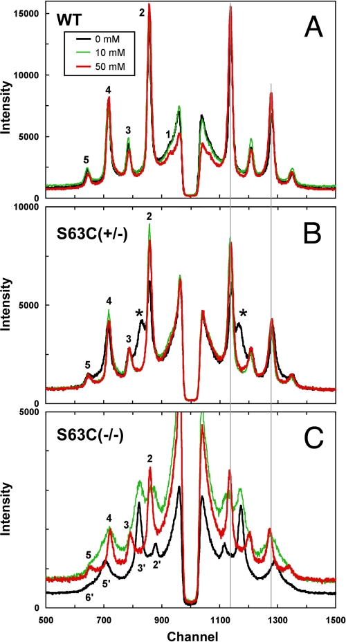
FIGURE 4.
Effect of reducing agent TCEP on swollen myelin in S63C peripheral nerve. Diffraction patterns from WT (A), S63C(+/−) (B), and S63C(−/−) (C) sciatic nerves treated for 15–20 h with 0, 10, or 50 mm TCEP in 154 mm NaCl, 5 mm Tris base, pH 7.4. When S63C(+/−) was treated with 10 mm TCEP, the swollen array disappeared revealing only the native 176-Å period structure. When S63C(−/−) was treated with 10 mm TCEP, two arrays were seen (187 and ∼222 Å), and after increasing the concentration of TCEP to 50 mm, only a single native period was observed. Bragg orders 2′, 3′, 5′, and 6′ index the swollen array, and 2–5 index the native array. The pair of thin vertical lines on the right-hand side that link the three panels indicate the positions of the 2nd and 4th order reflections for native period myelin.

