FIGURE 2.
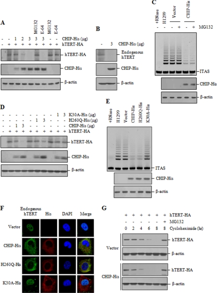
CHIP inhibits nuclear localization of hTERT and enhances hTERT degradation. A, H1299 cells were co-transfected with hTERT-HA and increasing amounts of CHIP-His and treated with or without 10 μm MG132 or E-64 for 2 h as indicated. The hTERT levels were measured by immunoblotting with anti-HA antibody. B, H1299 cells were transfected with or without CHIP-His, and lysates were analyzed by immunoblotting for the expression of endogenous hTERT and CHIP-His. C, H1299 cells transfected with CHIP-His or the empty vector were treated with or without 10 μm MG132 for 2 h, and lysates were analyzed for telomerase activity by the TRAP assay. D, H1299 cells were co-transfected with hTERT-HA and increasing amounts of CHIP-His, H260Q-His, or K30A-His. The hTERT levels were measured by immunoblotting with anti-HA antibody. E, H1299 cells transfected with CHIP-His, H260Q-His, or K30A-His were analyzed for telomerase activity by the TRAP assay. F, H1299 cells transfected with CHIP-His, H260Q-His, or K30A-His were treated with 10 μm MG132 for 2 h and subjected to indirect immunofluorescence with anti-hTERT (green) or anti-His (red) antibodies, followed by fluorescent microscopic observation. The nuclei were stained with 4,6-diamino-2-phenylindole (DAPI, blue). G, H1299 cells were co-transfected with hTERT-HA, along with CHIP-His or the empty vector, and treated with 100 μg/ml cycloheximide for the indicated times. Lysates were analyzed by immunoblotting with anti-HA or anti-actin antibodies. Cells were pretreated with 10 μm MG132 as indicated. ITAS represents the internal telomerase assay standard.

