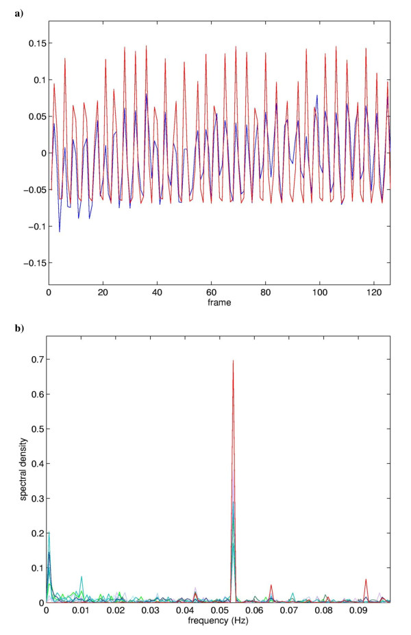
Figure 2.
Comparison of the mean temporal expression coefficient for MCI 1 with the modelled hemodynamic response (a) and the frequency power spectra of the underlying components temporal expression coefficients (b). Top: Time course of the average expression coefficient obtained from the eight PCs constituting MCI 1 (blue), superimposed on the hemodynamic model (red). The correlation coefficient between the time course and the model is 0.65. Bottom: Superposition of the frequency power spectra of the eight PCs constituting MCI 1, peak at about 0.054 Hz.

