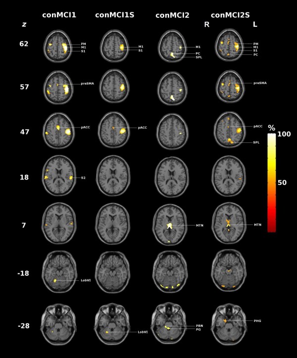
Figure 5.
Conjunction images of positively loaded regions (99th percentile) of task-related PCs. conMCI1, conMCI1S, conMCI2, conMCI2S : conjunction images (see text and Table 2 for details); MI: primary motor cortex, SI: primary somatosensory cortex, SII: secondary somatosensory cortex, PM: premotor cortex, PC: precuneus, SPL: superior parietal lobule, SMA: supplementary motor area, pACC: paralimbic anterior cingulate cortex, MTN: midline thalamic nuclei, PBN: parabrachial nucleus, PG: periventricular grey, LobVI: Cerebellum; Lobule VI, PHG: parahippocampal gyrus.

