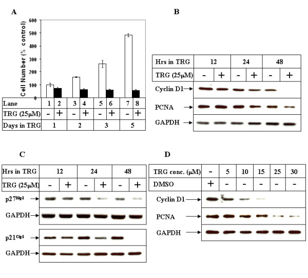
Figure 1.

Effect of TRG on HCC cell proliferation. (A) Subconfluent Huh-7 cells were plated on 6 well plates in regular growth medium. Next day, they were treated with either DMSO (-) or 25 μM TRG (+) and harvested at the indicated time intervals. The cell numbers were determined and represented as % control considering the DMSO-treated sample of 24 hours as 100%. Cells were plated in triplicate for each time point and each experiment was repeated at least twice. (B) Cells were treated as in A for the indicated time periods, following which they were harvested and total protein was extracted. Western Blot analysis of the cell extracts was then performed with antibodies against Cyclin D1, PCNA and GAPDH (as control). (C) The cells were treated as in B and cell extracts analyzed by Western Blots with the indicated antibodies. (D) Huh-7 cells were treated with either DMSO or increasing concentration of TRG for 48 hours, followed by Western Blot analysis with the indicated antibodies.
