
Figure 3.
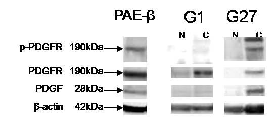
Protein levels of PDGF-B, PDGFR-β, and p-PDGFR-β evaluated by immunoblotting analysis in representative two cases (G1, PDGF-B overexpression -/p-PDGFR-β-; G27, PDGF-B overexpression +/p-PDGFR-β +) of tumor and paired normal tissue. Porcine aortic endothelial cells expressing PDGFR-β (PAE-β), was stimulated by PDGF-B and used as positive control for phosphorylation of PDGFR-β. N, normal tissue; C, cancer tissue.