
Figure 1.
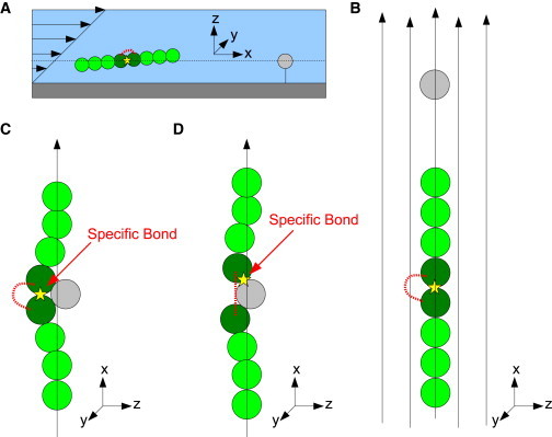
(A) Schematic of the flow chamber in our simulation. (B) Possible trajectories of the protein when moving in flow. (C) The masking domain is attached to the binding domain when the protein passes the ligand. The bond is denoted by a yellow star. (D) The masking domain is detached from the binding domain, and the binding domain forms a specific bond (yellow star) to the ligand. (Note that the protein length in this figure is arbitrary, and in most cases, the protein length is much longer than shown.)