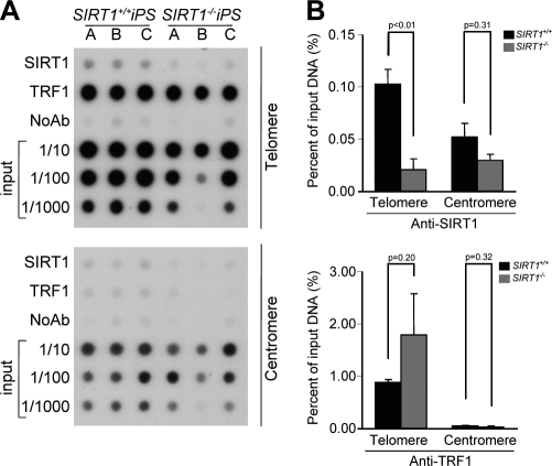
Figure 8.
SIRT1 protein interacts with telomeric repeats in vivo in iPS cells. (A) ChIP was performed with iPS cells of the indicated genotype (n = 3) using antibodies against SIRT1 and TRF1. Immunoprecipitated material was transferred to a nitrocellulose membrane and probed with a 1.6-kb telomeric probe (top) and a mouse major satellite (bottom). (B) Quantification of ChIP values for telomere and centromere repeats as indicated. The amount of immunoprecipitated DNA was normalized for the amount of telomere or centromeric repeats present in the cross-linked chromatin fraction unbound to the preimmune serum (input). n = number of independent MEFs used. Bars represent the mean between replicates, and SEM is shown. A Student’s t test was used to calculate statistical significance, and p-values are shown.

