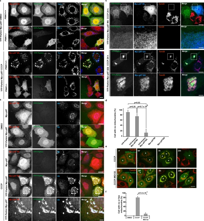
Figure 6.
p97 recruitment to ubiquitinated mitochondria mediates PINK1–Parkin-induced mitochondrial elimination. (a) MEFs from PINK1+/+ or PINK1−/− mice were transiently transfected with YFP-Parkin and Myc-p97. Cells were treated with DMSO or CCCP, then immunostained with anti-Myc and anti–cytochrome c. CCCP, 20 µM for 2 h. Bar, 10 µm. (b) HeLa cells transiently expressing Myc-p97 or YFP-Parkin/Myc-p97 were treated with DMSO or CCCP. Cells were immunostained with anti-Myc and anti-Ub (fk1). CCCP, 10 µM for 90 min. The boxed regions are magnified in the bottom panels. (c) HeLa cells were transiently transfected with YFP-Parkin and wild-type Myc-p97 or Myc-p97QQ. After treatment with CCCP for 24 h, cells were fixed and immunostained with anti-Myc and anti-Tom20 antibodies. CCCP, 10 µM. The boxed regions are magnified in the bottom panels. Bars: (top panels) 10 µm; (bottom enlarged panels) 1 µm. (d) Parkin-induced mitophagy in cells shown as in c was quantified (n > 50). CCCP, 10 µM for 24 h. Data represent the mean ± SD with P values of at least three replicates. (e) HeLa cells or HeLa cells expressing YFP-Parkin were treated with CCCP or CCCP plus MG132. Mitochondria were immunostained with anti-Tom20. Cells expressing YFP-Parkin are indicated with asterisks. CCCP, 10 µM for 24 h; MG132, 30 µM 30 min prior and with CCCP. Bar, 10 µm. (f) MG132 blocks Parkin-induced mitophagy in HeLa cells as in d. A quantification is shown. Data represent the mean ± SD with P values of at least three replicates.

