
Figure 3. Prolonged decay-time constants of NMDAR-mediated currents (EPSCs) post-lesion.
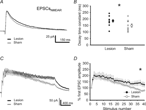
A, representative traces of NMDAR-mediated EPSCs recorded at a holding potential of +40 mV from lesion-treated (black trace) and sham-operated (grey trace) animals. B, the decay-time constants of these EPSCs were significantly prolonged in the vicinity of the lesion. C, representative current traces of NMDA-mediated EPSCs in response to high frequency presynaptic stimulation (40 pulses at 33 Hz). D, the mean relative amplitude of NMDAR-mediated EPSCs upon high-frequency synaptic stimulation was higher in the vicinity of the lesion as compared to sham-operated controls.