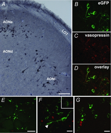
Figure 1. Vasopressin neurones in the AON.

Vasopressin neurones are found in all AON subdivisions in wild-type Sprague–Dawley rats (A; scale bar = 100 μm). eGFP-immunoreactivity mirrors vasopressin immunoreactivity in eGFP-vasopressin transgenic rats (B–D– AONl). BrDU labelling in the AON was typically found around the rostral migratory tract, while eGFP-vasopressin cells in the pars principalis were most often distributed in the superficial aspect of the deep cellular layer (Layer II). Vasopressin neurones were not co-localised with BrDU in the olfactory bulb (E and F– glomerular layer of the MOB; scale bar = 100 μm and 20 μm, respectively; F– arrow shows location of orthogonal view in the boxed inset) or in any region of the AON (G– lateral AONl; scale bar = 20 μm), suggesting that vasopressin cells in the AON are not simply newly born neurones.
