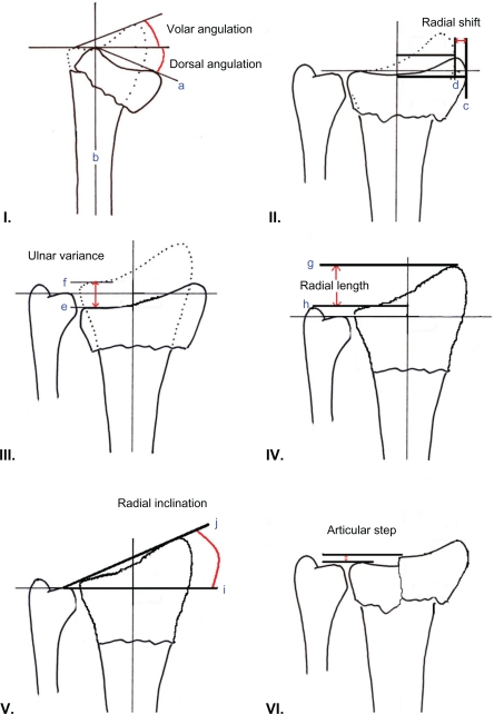
Figure 1.
Radiographic measures of outcome in distal radius fractures. I) Dorsal angulation: The angle between the line which connects the most distal points of the dorsal and volar cortical rims of the radius (a) and the line drawn perpendicular to the longitudinal axis of the radius (b). Normally 11°–12° volar. II) Radial shift: This is a relative measurement, which is taken as the difference between the measurements of the fractured radius (c) and the normal, uninjured radius (d). III) Ulnar variance: Vertical distance between a line drawn parallel to the proximal surface of the lunate facet of the distal radius (e) and a line parallel to the articular surface of the ulnar head (f). Usually negative variance −1 mm. IV) Radial length: Distance between a line drawn at the tip of the radial styloid process, perpendicular to the longitudinal axis of the radius (g) and a second perpendicular line at the level of the distal articular surface of the ulnar head (h). Normally 11–12 mm. V) Radial inclination: Angle between a line perpendicular to the longitudinal axis of the radius (i) and a line joining the distal tip of the radial styloid and the distal sigmoid notch (j). Usually 21°–25°. VI) Articular step: Up to 2 mm is acceptable.

