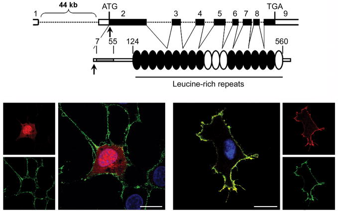
Figure 7. The disease-causing 4A > G (Ser2Gly) change in SHOC2 promotes protein myristoylation and cell membrane targeting.
(A) SHOC2 genomic organization and protein structure. The coding exons are shown at the top as numbered filled boxes. Intronic regions are reported as dotted lines. SHOC2 motifs comprise an N-terminal lysine-rich region (grey coloured) followed by 19 leucine-rich repeats. Numbers above the domain structure indicate the amino acid boundaries of those domains. (B) Confocal laser scanning microscopy analysis documents that SHOC2wt (red) is uniformly distributed in the cytoplasm and nucleus (left) of transiently transfected Cos-1 cells (DMEM supplemented with 10% heat-inactivated FBS), while SHOC2S2G (red) co-localizes with ganglioside M1 (green) to the cell membrane.39 Nuclei are visualized by DAPI staining (blue). Bars indicate 20μm.

