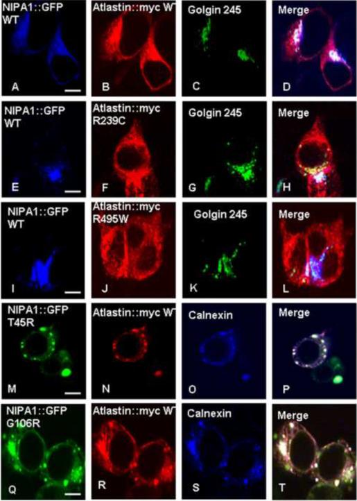
Figure 2. The presence of mutant forms of binding partners caused sequestration of WT NIPA1 in GC and WT atlastin-1 in ER.
Triple immunostaining confirming the colocalization in GC complex of both WT forms of proteins (panels A–D) and in ER (panels A–D, calnexin co-staining not shown). We also observed an accumulation of WT NIPA1 in GC in the presence of studied HSP atlastin-1 mutations (panels E–L), and accumulation of WT atlastin-1 in ER together with mutant forms of NIPA1 (panels M–U). Scale bar = 5μm.

