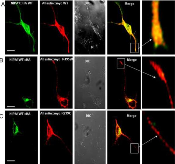
Figure 8. Overexpression of WT NIPA1 did not compensate for the effect of mutant forms of atlastin-1.
Overexpression of WT NIPA1 did not alter the patters illustrated in Figure 7 and the sequestration of WT NIPA1 in the neuronal bodies was again seen after the introduction of HSP atlastin-1 mutations. Panel A shows distribution of co-expressed WT NIPA1-HA and WT atlastin-1-myc proteins (green anti-HA antibodies, red anti-myc antibodies). Panels B (expressed mutation R495W atlastin-1-myc) and C (expressed mutation R239C atlastin-1-myc) demonstrate markedly altered intracellular distribution of expressed WT NIPA1 with its absence from axons. Scale bar = 10μm.

