
Figure 4. CpG ODN accelerate neovascularization of biopsy sites.
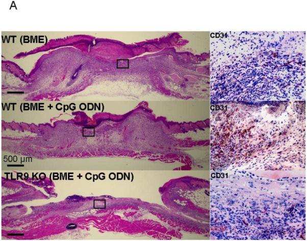
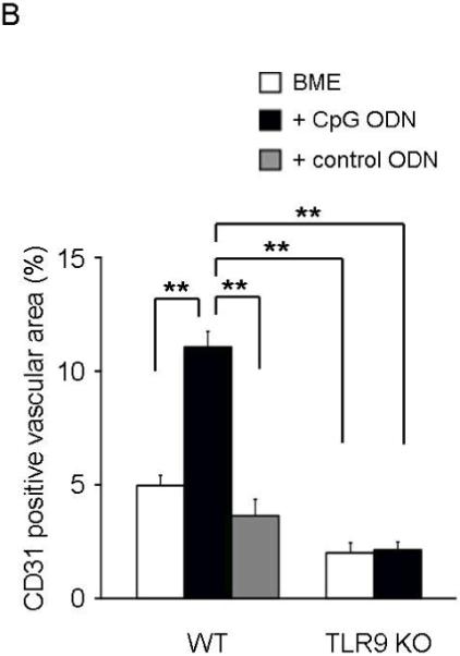
Histologic sections were obtained from biopsy sites as described in Fig 3. (A) Photomicrographs of representative wild type (upper and middle panels) and TLR9 KO mice (lower panel; original magnification 20X). The insets show higher magnifications (400X) of CD31-stained sections of granulation tissue. (B) The re-vascularized area was calculated based on CD31 staining of representative tissue sections using Image J. Results represent the mean + SE of 2-3 independent experiments with 4-7 animals/group.
**, p < 0.01.