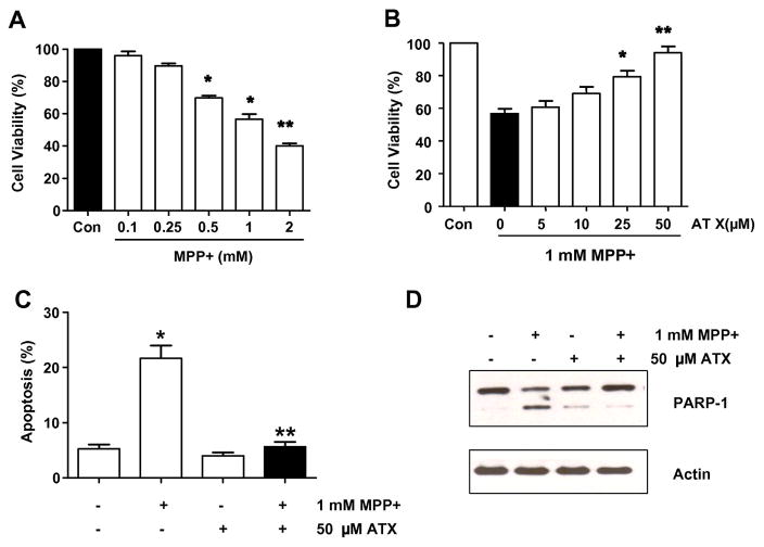
Fig. 2.
Effect of AST on the MPP+-induced decrease in SH-SY5Y cell viability. Cell viability (A and B) or apoptotic death (C and D) was assessed by MTT assay, flow cytometric assay, or immunoblotting assay as described in the Materials and Methods section. (A) Cells were treated with various concentrations (0.1–2 mM) of MPP+ for 24 h. (B) Cells were pretreated with various concentrations (5–50 μM) of AST for 2 h and then treated with 1 mM MPP+ for 24 h in the presence of AST. (C) Cells were treated with either 0.1% DMSO (control) or 1 mM MPP+ in the absence or presence of 50 μM AST for 24 h as described in B. After treatment, cells were stained with fluorescein isothiocyanate (FITC)-Annexin V and propidium iodide (PI). Apoptosis was detected by the flow cytometric assay. In A, B, and C, error bars represent the mean ± SE from three separate experiments. *p < 0.05, compared with control (untreated group); **p < 0.01, compared with the group treated by MPP+ alone. (D) Cells were treated as described C and then harvested. Lysates containing equal amounts of protein (20 μg) were separated by SDS-PAGE and immunoblotted with anti-PARP-1 antibody. Actin was shown as an internal standard.

