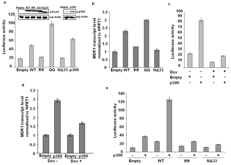
Figure 4.
Enhancement of MDR1 promoter activity and expression by APE1 acetylation. (a) HEK-293T cells were cotransfected with MDR1 promoter-luciferase reporter plasmid and empty vector, WT, RR, QQ, NΔ33 APE1 or p300 expression plasmids. 48 hours later, luciferase activity was measured and normalized with total protein content in the lysates. Inset panel shows the level of FLAG tagged WT/ mutant APE1 proteins or p300 protein in the cell extracts determined by Western analysis. (b) Real Time RT-PCR assay for MDR1 transcript levels in HEK-293T cells ectopically expressing WT, RR, QQ or NΔ33 APE1 mutant proteins. Values in the bar diagram are relative to empty vector transfected cells. (c) APE1 levels in APE1siRNAHEK-293T cells were downregulated with Dox treatment for 8 days. Then the cells were cotransfected with MDR1 promoter reporter plasmid and expression plasmids for p300 or its empty vector. Luciferase activity was measured as mentioned above. (d) MDR1 transcript levels in Dox-treated (APE1 downregulated) or untreated APE1siRNAHEK-293T cells ectopically expressing p300. Values in the bar diagram are relative to empty vector transfected cells. (e) HEK-293T cells were cotransfected with MDR1 promoter reporter plasmid along with WT, RR or NΔ33 APE1 constructs and p300 expression plasmid. Luciferase activity was measured as mentioned above. Results in all the cases represent the mean ± standard deviations of 3 independent experiments performed in duplicates.

