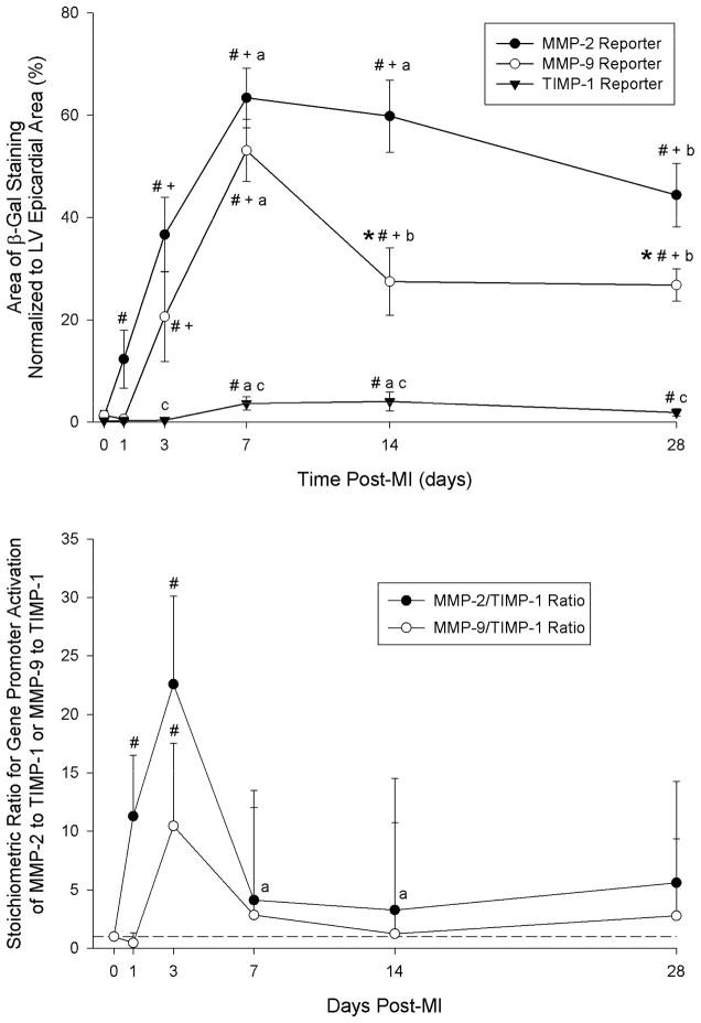
Figure 2.
TOP: Summary data for area of positive β-galactosidase staining normalized to LV epicardial area. Sample sizes for each group at each post-MI time point are presented in Table 1. # p<0.05 vs. Acute (1 hour post-MI), + p<0.05 vs. 1 day post-MI, a p<0.05 vs. 3 days post-MI, b p<0.05 vs. 7 days post-MI, *p<0.05 vs. MMP-2 Reporter values only, c p<0.05 vs. MMP-2 and MMP-9 Reporter values. BOTTOM: Ratios of MMP-2 to TIMP-1 promoter activation and MMP-9 to TIMP-1 promoter activation at each post-MI timepoint. These ratios were computed as a function of the average β-galactosidase staining recorded in the TIMP-1 reporter group at each respective post-MI timepoint. The maximum change in MMP-2/TIMP-1 or MMP-9/TIMP-1 ratios occurred at 3 days post-MI and was normalized at later post-MI durations. # p<0.05 vs. Acute (1 hour post-MI), + p<0.05 vs. 1 day post-MI, a p<0.05 vs. 3 days post-MI.

