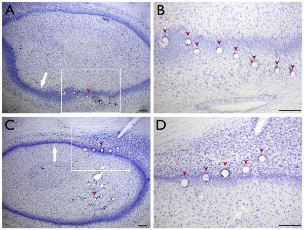
Figure 1. Histological confirmation of electrode location.
(A, C) Nissl-stained transverse sections through the hippocampus in two animals. The tissue damage due to electrode placement (red arrowheads) can be seen along the pyramidal cell layer (white arrow). (B, D) Higher magnification of the boxed portions of (A) and (C), respectively. All scale bars 100μm.

