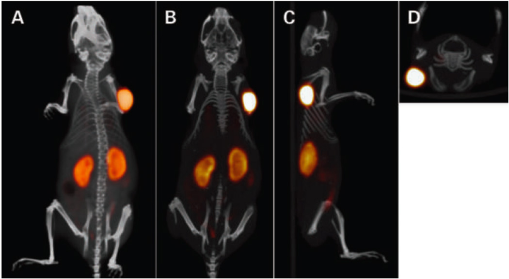
Figure 3.
Static PET/CT imaging study of a BALB/c nude mouse with LS174T tumor (0.1 g) on the right side, which received 6.0 nmol TF2 and 0.25 nmol 18F-IMP-449 (5 MBq) intravenously with a 16-hr interval [6••]. The animal was imaged 1 hr after injection of 18F-IMP-449. The panel shows the three-dimensional volume rendering (posterior view; A) and cross-sections at the tumor region: coronal (B), sagittal (C), and transverse (D). Credit: Schoffelen R, Sharkey RM, Goldenberg DM, Franssen G, McBride WJ, Rossi EA, Chang C-H, Laverman P, Disselhorst JA, Eek A, et al.: Pretargeted Immuno-Positron Emission Tomography Imaging of Carcinoembryonic Antigen-Expressing Tumors with a Bispecific Antibody and a 68Ga- and 18F-Labeled Hapten Peptide in Mice with Human Tumor Xenografts. Molecular Cancer Therapeutics 9:1019–1027. ©2010 American Association for Cancer Research. Adapted with permission.

