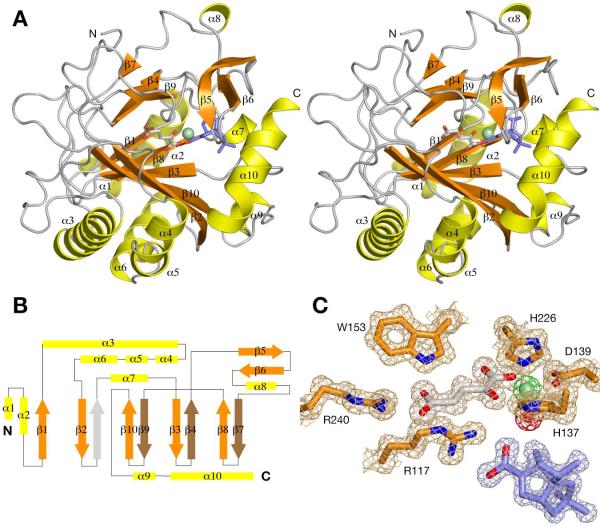
Figure 3.
Structure of PtlH
(A) Stereo diagram showing a schematic representation of the structure of PtlH. The Fe(II) ion is shown as a light-green sphere, α-ketoglutarate is shown as sticks with carbon atoms in gray, and ent-1PL is shown as sticks with carbon atoms in blue. (B) Topology diagram of PtlH. The position of the β-strand not present in the PtlH DSBH fold is indicated with a gray arrow. (C) Final 2Fo-Fc electron density map (contoured at 1σ) of the active site region. Electron density for Fe(II), α-ketoglutarate, ent-1PL and the Fe(II)-coordinated solvent water molecule is shown in green, gray, blue and red, respectively.

