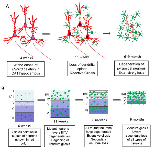
Figure 11. Schematic models of pathologic changes in the mutant brain over times.
A. Schematic drawing depicts the temporal phenotype of the CA1 hippocampal neuron in the mutant brain. Hippocampal neurons in the CA1 region appear normal at 4 weeks when Pik3c3 deletion occurs. Seven weeks later, hippocampal neurons show loss of dendritic spines accompanied with apparent gliosis. At late stage, extensive pyramidal neuronal degeneration and gliosis occur.
B. Schematic model summarizes phenotypes observed in the mutant cortex. A small subset of cortical neurons undergo CamKII-Cre mediated Pik3c3 deletion, and the cortex appears normal at 4~6 weeks. Layer III and IV mutant cortical neurons degenerate which is associated with reactive gliosis at 11 weeks. At 6 months, all Pik3c3 deficient cortical neurons degenerate, some loss of wildtype neurons begins and extensive gliosis occurs. At late stage, severe loss of all types neurons occurs, concurrent with extensive gliosis that covers the remaining cortex.

