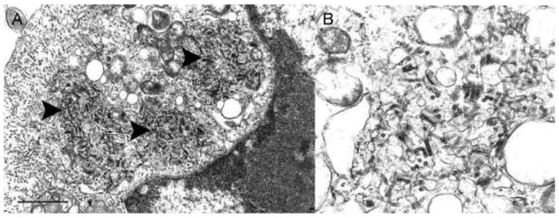
Fig. 2.

Ultrastructure of DURV in ultrathin sections of Vero cells. (A) Portion of a cell cytoplasm with three areas of virus formation (arrowheads); Bar = 1 μm. (B) Detail of a virus formation area, demonstrating virions budding into enlarged endoplasmic reticulum cisterns; Bar = 100 nm.
