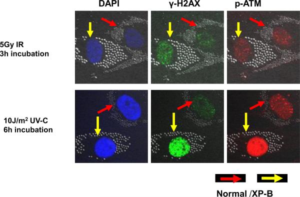
Figure 1.
Immunofluorescence of phosphorylated histone H2AX and ATM after uniform ionizing radiation (IR) compared to uniform UV in normal and XP-B (XP183MA) cells. Normal (0.8 μm beads – red arrows) and XP-B cells (2.0 μm beads- yellow arrows) on the same slide were exposed to 5 Gy IR (top row) or 10 J/m2 UV (bottom row) and then cultured for 3 h or 6 h before fixation. Immunofluorescent double labeling revealed that IR and UV irradiation led to phosphorylation of the DSB-related proteins H2AX and ATM in the normal and XP-B cells.

