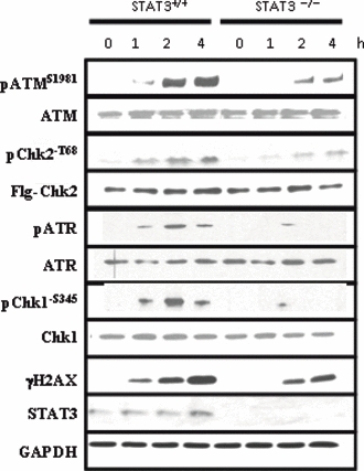
Figure 2.

Phosphorylation of ATM and H2AX are reduced in STAT3−/− MEFs. STAT3+/+ and STAT3−/− MEFs were treated with DMSO (0 h) or 10 μM etoposide for 1, 2 or 4 h and western blots were carried out on cell lysates using the indicated antibodies for ATM, phospo-ATM (pATMS1981), Chk2, phospho-Chk2 (pChk2-T68), ATR, phosphor-ATR (pATR), Chk1, phosphor-Chk1 (pChk1-S345), γH2AX, STAT3 and GAPDH. For Chk2 and pChk2-T68 immunoblots, cells were transfected with human Flag-tagged Chk2 (1μg/time point) and Western blotted with the specific anti-Flag antibody to measure the levels of transfected Chk2 (Flg-Chk2) or an anti-pChk2-T68 following etoposide treatment.
