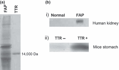
Figure 1.

(a) SDS-PAGE silver stain of FAP human kidney aggregate extracts and TTR standard; the αB-crystallin band is pointed out by a rectangle. (b) αB-crystallin western blot of i) human normal and FAP kidney; ii) transgenic mice stomach aggregate extracts (n = 3 each) with and without TTR deposition (TTR+ and TTR− respectively)
