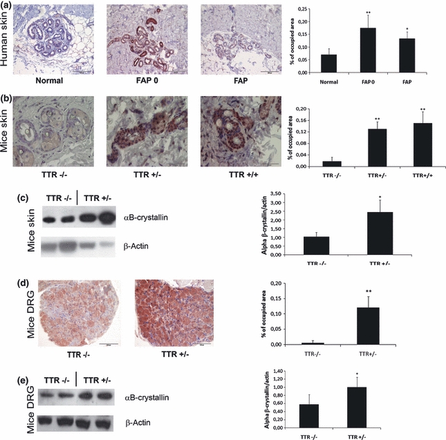
Figure 3.

TTR deposition induces αB-crystallin expression. (a) immunohistochemistry of human skin, showing increased expression in FAP 0 (n = 5) and FAP patients (n = 3) when compared to controls (n = 5) (bar = 200 μm); (b) immunohistochemistry of transgenic mice skin: increased expression of αB-crystallin in TTR+/− (n = 4) and TTR+/+ (n = 4) transgenic mice when compared to mice without TTR deposits, TTR−/− (n = 5) (bar = 50 μm); (c) western blot of transgenic mice skin for αB-crystallin: increased expression of αB-crystallin in animals with TTR deposits (TTR+/−, n = 4) when compared with animals without TTR deposits (TTR−/−, n = 4); (d) immunohistochemistry of hTTR HSF1-het transgenic mice DRG, showing increased expression of αB-crystallin in mice with TTR deposits (TTR+/−, n = 4) when compared to mice without TTR deposition (TTR−/−, n = 4) (bar = 100μm); (e) Western blot analysis of αB-crystallin expression in DRG of hTTR HSF1-het showing increased expression of αB-crystallin in mice with TTR deposits (TTR+/−, n = 7) when compared to mice without TTR deposition (TTR−/−, n = 7). Quantification of images and blots are shown on the charts on the right (*P < 0.01, **P < 0.005).
